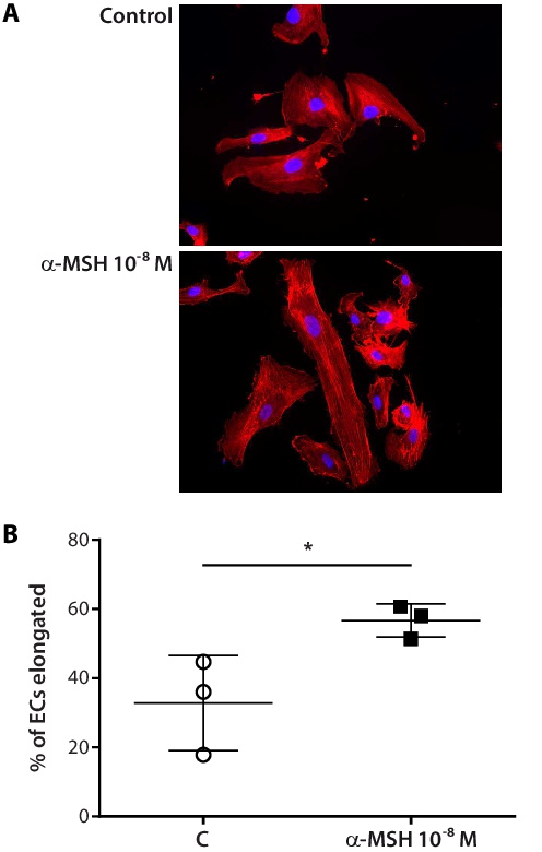
Fig. 3. MC1R activation enhances actin filament remodelling and cell elongation in migrating HAoECs. (A) Non-confluent ECs were stimulated with α-MSH for 6 h, then fixed and stained with TRITC-labelled phalloidin for actin filament visualization, using DAPI for nuclear counterstain (40×). Aligned stress fibres and cellular elongation are pronounced in treated vs. untreated HAoECs. (B) Quantification of cell elongation. Cells with axial ratios > 3 were counted in randomly selected fields and expressed as percentages of the total cells counted. Results are shown as scatter dot plots with mean ± SD (n = 3 per group). Statistical significance of differences was assessed by two-tailed unpaired t test (*p=0.0356).